
Oral Lichen Planus: What You Need to Know
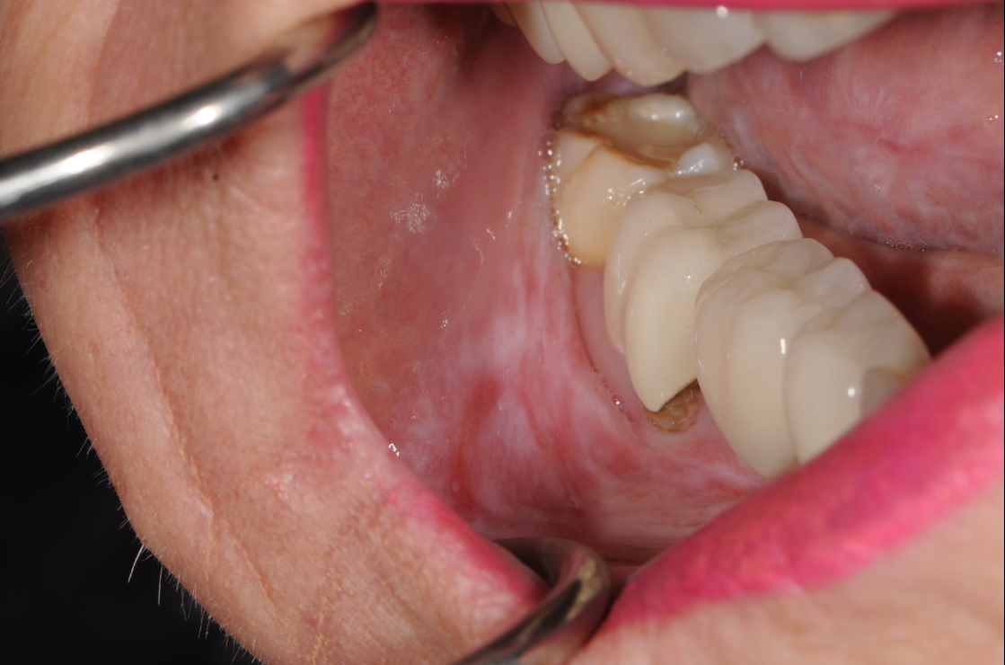
Clinical photo of oral lichen planus showing characteristic white, lacy striations (Wickham’s striae) on the inner cheek. Common presentation of chronic inflammatory oral mucosal disease. Useful for identifying symptoms of oral lichen planus in dental and medical diagnostics. CJ Henley, DMD, PA – Jacksonville, FL – for educational purposes on oral lichen planus diagnosis and treatment.
Oral lichen planus is a chronic inflammatory condition that affects the soft tissues inside the mouth. It often appears as white, lacy patches or red, swollen areas, and can cause burning or discomfort, especially when eating spicy or acidic foods. While not contagious or cancerous, it requires proper diagnosis and regular monitoring. At CJ Henley, DMD, PA in Jacksonville, FL, we offer personalized treatment for oral lichen planus to help manage symptoms and protect your oral health.
-
Description text goes hereWhite, lacy patches inside the cheeks or on the tongue
Red, swollen tissues
Burning or pain, especially with spicy or acidic foods
Sores that may come and go
If you’re experiencing these symptoms, early evaluation is key.
-
The exact cause is unknown, but it’s believed to be related to the immune system. It may be triggered or worsened by:
Stress
Certain medications (like NSAIDs or beta-blockers)
Dental materials (such as amalgam)
Infections like hepatitis C (rarely)
-
Treatment focuses on managing symptoms and preventing flare-ups. At CJ Henley, DMD, PA, we tailor your care plan to your needs. Options include:
Biopsy to determine proper diagnosis
Good oral hygiene and gentle dental care
Topical corticosteroids to reduce inflammation
Anti-inflammatory rinses or gels
Regular monitoring for changes (especially if lesions are erosive)
-
Avoid spicy, acidic, or rough-textured foods
Quit tobacco products and reduce alcohol intake
Reduce stress when possible
Use gentle toothpaste (free of sodium lauryl sulfate)
-
What Oral Lichen Planus Looks Like Under the Microscope (Histology)
To accurately diagnose oral lichen planus (OLP), we sometimes use a small tissue sample (biopsy) to examine the area under a microscope. This helps us distinguish it from other similar conditions, such as leukoplakia, lupus, or oral cancer.
Key Histological Features of Oral Lichen Planus:
Saw-tooth rete ridges: The interface between the epithelium and connective tissue shows a jagged, saw-tooth appearance.
Band-like lymphocytic infiltrate: A dense collection of immune cells (mainly T-lymphocytes) hugs the basement membrane zone, causing inflammation.
Basal cell degeneration: The basal layer of the epithelium shows damage or destruction, often described as liquefaction degeneration.
Civatte bodies: These are degenerated keratinocytes (epithelial cells) found in the lower layers of the epithelium or in the connective tissue.
These findings confirm the immune-mediated nature of the disease and help guide treatment.
-